Blog Hero

Why Does the Optometrist Take Pictures of My Eyes?

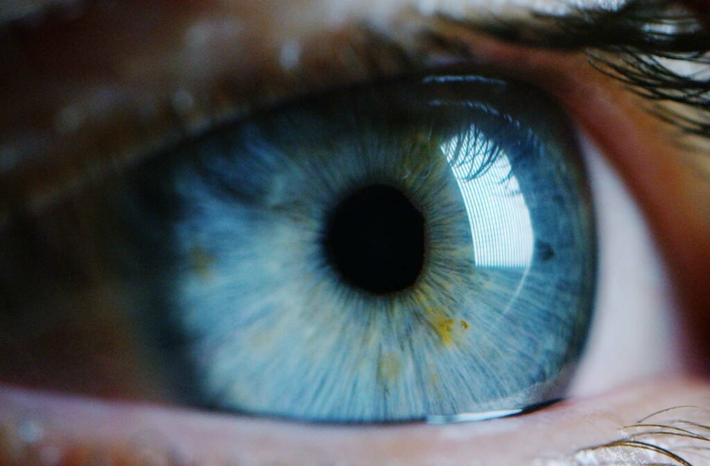
Fundus photography, or a picture of the inside of the eye, is an important part of your comprehensive eye examination.  

The camera used is designed to allow high-resolution photography of the retina and the optic nerve.  Although the optometrist is able to view these structures of the eye with a microscope and lens during your examination, the purpose of the photo is threefold:

  1. it serves as documentation (photo-documentation) of the health of the inside of your eye at that specific time, allowing for comparison at future visits
  2. it helps in the identification and diagnosis of disease and abnormalities involving your retina and optic nerve, like glaucoma, macular degeneration, and changes due to diabetes to name a few
  3. it allows the optometrist to educate you about your eye health, specifically by showing  you any issues visible on the photo.

Written by Dr. Serge Fauchon

After studying physics at the University of Ottawa, Dr. Fauchon graduated from the University of Montreal, School of Optometry in 1997.

He did an internship at the Quentin Burdick Memorial Hospital in Belcourt, North Dakota, with an emphasis on diseases of the retina and glaucoma.

Dr. Fauchon joined eyeDOCS in 1997 and became partner in 1998.

He provides full-scope optometric care with a special interest in eye disease, particularly for glaucoma and diabetic patients. He also works closely with laser centers for pre- and post-refractive laser surgery co-management. He enjoys going to different schools in the Ottawa area to perform eye exams for junior and senior kindergarten students.

More Articles By Dr. Serge Fauchon
instagram facebook facebook2 pinterest twitter google-plus google linkedin2 yelp youtube phone location calendar share2 link star-full star star-half chevron-right chevron-left chevron-down chevron-up envelope fax